The necessity for a dental bone graft depends on the jaw bone anatomy. Replacing missing bone or adding mass to existing bone is often essential to the success of a dental implant. An implant needs a critical mass of bone surrounding it in order to bind (osseointgrate) to it and deliver sufficient strength and stability.
A key to dental implant success is the amount and quality of the bone where the implant is to be placed. Ridge deformities of the upper and lower jaw can leave you with inadequate bone and tissue thickness for an esthetic dental implant restoration. The defects may have been caused by trauma, developmental defects, periodontal disease and wearing dentures.
If teeth have been missing for some time, the ridge will shrink in both height and width. In severe cases, the ridge will not be wide enough or high enough to place a dental implant. A bone graft is placed to augment ridge height and/or width to provide proper anchorage to surround the implant with healthy strong bone. After sufficient healing (6-8 months) the implants are placed during a second surgical procedure.
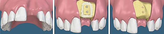
Pictures demonstrate block bone grafting procedure
This technique involves mechanical expansion of the bony ridge. This is a technique used to restore the lost bone dimension when the jaw ridge gets too thin to place dental implants. In this procedure, the bony ridge of the jaw is literally expanded by mechanical means at the time the actual implant is placed. Bone graft material can be placed to supplement the thickness of the ridge around the implant. This is not helpful for increasing the bony height of dental ridges, only the width.
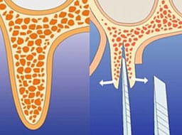
Pictures demonstrate ridge expansion in case which the jaw bone width is >4mm.
A socket is the name for the area in the bone in which a tooth is rooted. When a tooth is extracted or previously lost, the soft tissue and bone collapse. Furthermore, if a socket remains empty after a tooth is lost or extracted the accelerated bone loss in the area can also negatively impact any adjacent teeth. Therefore, it is very important for health and esthetic to rebuild the socket as soon as possible after tooth loss or extraction. The dental surgery done to prevent this area from collapsing is called ridge or socket preservation in order to allow tooth replacement by implant or bridge restoration. A ridge preservation surgery can serve many purposes. It is most useful in preserving the natural appearance of the front of the mouth. It is also very instrumental in providing appropriate support for dentures or dental bridge construction. It can be essential in providing sufficient bone height to support dental implants. Dr. Hanasab recognizes the role of ridge preservation in providing the support for more natural, appealing and functional tooth replacement.
Socket preservation will begin with the removal of a tooth if it has not already been lost. Any remaining root particles will be removed to leave a clean empty socket.
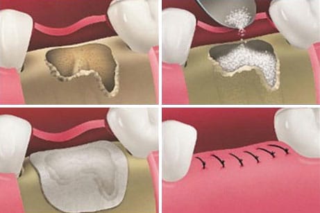
Material will then be placed into that socket to fill it and build it into a firm foundation for reconstruction. A guided bone regeneration membrane is placed over the grafted material. There will usually be an extended healing time for the grafted bone to fully integrate with the existing natural bone. It is important to wait until this healing process is truly complete before continuing with the restoration in the area. When the new bone is well fixed then implants or bridgework can restore full oral function. Socket and other types of bone grafting have allowed the placement of implants for patients who would otherwise not be candidates for this type of restoration.
Autogenous - meaning using the patients own bone that is taken from other areas in the mouth (chin, ramus, or tuberosity : see a picture below) as the dentist collects it while he drills into the bone to prepare the site for the dental implants. In some rare cases bone is taken from areas outside the mouth (hip) – but that procedures is preformed at a hospital. This type of bone grafting is expected to give the best results for jaw augmentation.
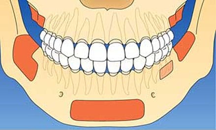
Block Bone Grafting: Donor Areas
Alloplast - synthetic bone. While using this type the dentist mixes in the patient's blood to help the material accelerate and promote bone formation in the graft area.
Xenograft - bone taken from cow. This bone is harvested under very strict supervision and it is very safe. After applying the bone grafting materials, a collagenmembrane is placed to hold the material in place. The membrane holds the material in place preventing soft tissue to blend in, enabling the material to regenerate and form new bone. This type of grafting material has very good result for sinus grafting procedure.


Pattaya City: One of the hottest beach-resort destinations in Thailand, Pattaya may not be idyllic but it certainly
makes up for it with a wide variety of activities, accommodation, and nightlife venues. Read more
OrthoSmile is a full service dental clinic in Pataya offering a comforting and relaxing atmosphere one finds in an upscale day spa. Drs. Kasidis, Raungyos and Porndee apply their leading dental expertise in this relaxing retreat, leaving clients refreshed, healthy and rejuvenated.
- Vistascan dental x-ray scanner
- Consult Pro dental software
- Osim Udivine
- LaserSmile
- Invisalign
- Anthos dental chair
- Ostell mentor
- Piezosurgery
Phone:
(038)488354
(038)488355
(089)6976943
Ortho Smile Dental 382/9-10 M9 Pattaya 2nd Rd. Soi 6/1 Pattaya City Banglamung Chonburi Email : [email protected]
Tel : 038 488 353-5 All Right Reserved © 2010 www.Orthosmiledental.com Designed by Cybrilliant.com